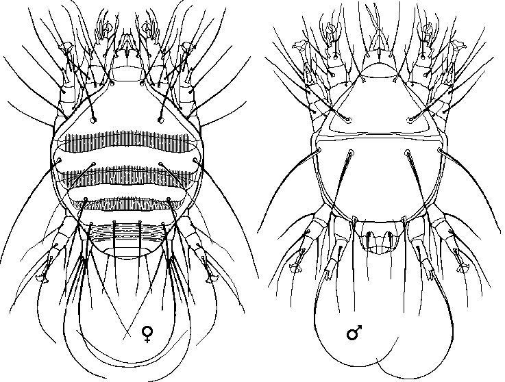
 The mites can to be observed inside the tracheae or removed from them to be observed independantly using a microscope or high power hand lens.
The thoraces of suspect bees can be dissected to expose the trachea. Each trachea is examined using a microscope, the mites can be seen through the transparent wall of the trachea.
Larger samples of suspect bees can be ground up or homogenised in water, followed by coarse filtration of the suspension, and centrifugation. The deposit is treated with undiluted lactic acid for 10 minutes and mounted for microscopic examination. This method is not discussed further here as it is quite complicated for the amateur beekeeper.
Acarine mites can be stained so that they can be observed in stronger contrast within the bee trachea.
The honey bee problem known as Acariosis or Acarine, often called a disease, is actually an infestation of adult bees of Apis mellifera and other Apis species, caused by the microscopic Tarsonemid mite Acarapis woodii. In USA they are known as tracheal mites.
The Acarine mite is approximately 150 μm in size, and it is an internal parasite of the respiratory system of honey bees. The mites enter, live, and reproduce mainly in the large prothoracic tracheae, feeding on the haemolymph of their host. Sometimes, particularly in strong infestations, they may also be found in the head, thoracic and abdominal air sacs.
The symptoms found in infected bees depend on the number of parasites within the trachea and are due to physical injuries and to physiological disorders that are caused by the obstruction of the airways, lesions in the walls of the trachea, and to a minor extent depletion of haemolymph. As the number of mites increases, the walls of the trachea change from white/translucent to opaque and discoloured with irregular blotchy dark patches.
The infestation spreads by direct contact, only newly emerged bees less than 10 days old are susceptible. Mite reproduction occurs within the tracheae of adult bees, where female acarine mites may lay between eight and twenty eggs. There are two to four times as many females as there are males. Development takes 11 or 12 days for males and 14 or 15 days for females. Light infestations occur near to the spiracle opening and heavier infestations reach deeper into the tracheal network.
There are no reliable external clinical signs for the diagnosis of acariosis as the signs of affliction are not specific and the bees behave in much the same way as bees that are affected by various maladies. They crawl around on the ground in front of the hive and climb blades of grass, unable to fly. Dysentery and/or signs of 'K' wing may be present.
Identification of the problemSome claim that Acarine infestation can only be detected under laboratory conditions using microscopic examination or an enzyme-linked immunosorbent assay (ELISA). There is no reliable method for detection of very low levels of infection.
The number of bees sampled determines the detection threshold of the method. It has been shown that a 2% rate of infection can be detected by sampling 50 bees, while a 1% rate of infection is detected using 100 bees (confidence limit is 80% for a colony of average size in spring). Because of the high level of manual work involved, it is suitable to examine 50 bees. Sequential sampling is a neat trick to reduce the overall workload.
 Sequential Testing
Take a sample of 50 bees kill them then and examine them one by one. You plot the result on the graph depending whether they are positive or negative. The graph is basically cumulative positive versus No. of bees examined.
Once the plot gets above the upper line the sample has an infection level above the probability selected (in the case of diagram it is 20%), if the plot lies between the two lines, then further bees need to be examined, and if the plot dips below the lower line then the level of infestation is below the lower limit set for action. (Original material for the diagrams, supplied by Ruary Rudd.)
 Dissection to expose Acarine infestation
A random sample of 50 bees is collected from the suspect colony. These should be mainly bees crawling and unable to fly, found within about 3 metres of the front of the hive, rather than random collection from within the colony. The sample bees may be living, dying, or dead. Live bees must first be killed with ethyl alcohol or in a deep freeze at -20°C.
Each bee should be impaled, using a double needle placed at an angle away from the head through the thorax between the second and third pairs of legs (as shown at right). The bee should be ventral side up on an angled cork base, the angle is not critical, but is usually between 45° and 60°. Using a single edged razor blade, cut off the head and first pair of legs, the cut should be made from behind the first pair of legs to the back of the bee's head, indicated by the red line on the drawing, the severed head and front pair of legs can then be removed using tweezers.

Fine tipped tweezers can be used to peel away the collar (shown red at right) in order to expose the tracheae more fully. Pull upwards with a circular motion, following the ring of the collar. It will peel off easily, usually in one piece. The collar itself can be saved for later preparation as a microscope slide specimen, if required, by immersing in 70% isopropyl alcohol.

As mites enter through the spiracle check the outer end of the trachea first. Light infestations may be difficult to see, heavy infestations are easily visible as shadows or lumpy dark objects in trachea that can be clear to dark brown. Old and/or heavy infestations will render the trachea orange, brown or black.
Alternative Method
The trachea can be isolated by cutting a disc through the thorax in front of the middle pair of legs and the base of the forewings using a razorblade. These thin disks can then be further treated to clear muscle tissue by macerating in a 10% solution of potassium or sodium hydroxide boiling for a few minutes or by leaving them to stand overnight at room temperature until the soft internal tissues are dissolved and cleared leaving the chitinous parts intact. Using heat is more reliable as you can constantly monitor progress. Wash the sections in tap water to remove the alkali.
Examine the main pair of trachea using a dissecting microcope or a stereo microscope that gives an erect image. A magnification of x18 or x20 is suitable, or the trachea can be cut from the chitin ring and transferred to a glass slide, add glycerin or water and observe at whatever magnification you choose.
At low magnifications the mites are visible through the transparent tracheal wall, but are small, indistinct oval bodies, with higher magnifications the mites can be recognised.
Preferential StainingThe mites and trachea can be stained specifically, rendering them more easily visible under the microscope.
Remove the head and forelegs, create thoracic discs 1 mm to 1.5 mm thick, then clear the sections using a 10% solution of potassium or sodium hydroxide and wash as described above.
Staining and mount the sections... Cationic stains are the most suitable and specific as they stain the mites more intensely than they stain the trachea containing them. A solution of 1% aqueous methylene blue is the most suitable staining agent and it can be prepared by dissolving the methylene blue first and then adding sodium chloride to make a 0.85% or 0.9% NaCl solution. To actually stain the specimens, immerse in 1% aqueous methylene blue solution for 5 minutes differentiate sections in distilled water for 2 to 5 minutes, rinse the sections in 70% alcohol (ethyl or isopropyl). Examine the disks under a dissecting microscope at any appropriate magnification (10x up to 30x).
ReferencesBancroft J.D. & Stevens A. (1982). Theory and Practice of Histological Techniques. Churchill Livingstone, Edinburgh, UK.
Colin M.A., Faucon J.P., Gianfert A. & Sarrazin C. (1979). A new technique for the diagnosis of Acarine infestation in honey bees. Journal of Apicultural Research, 18, 222-224.
Grant G., Nelson D., Olsen P. & Rice W.A. (1993). The ELISA detection of tracheal mites in whole honey bee samples. American Bee Journal, 133, 652-655.
Peng Y. & Nasr M.E. (1985). Detection of honey bee tracheal mites Acarapis woodi by simple staining techniques. Journal of Invertebrate Pathology, 46, 325-331.
Mozes-Koch R. & Gerson U. (1997). Guanine visualization, a new method for diagnosing tracheal mite infestation of honey bees. Apidologie, 28, 3-9.
Wolfgang Ritter (1996). Diagnostik und BekÄmpfung der Bienenkrankheiten (Diagnosis and control of bee diseases). Gustav Fischer Verlag, Jena, Stuttgart, Germany.
FAKHIMZADEH, K. 2001. Detection of major mite pests of Apis mellifera anddevelopment of non-chemical control of varroasis. ? Doctoral Dissertation, Universityof Helsinki, Department of Applied Biology, Publication no: 3, Helsinki, 46 pp. +appendix articles. ISBN 951-45-9914-4.
Sammataro D. & Needham G.R. (1996). Host-seeking behaviour of tracheal mites (Acari: Tarsonemidae) on honey bees (Hymenoptera: Apidae). Experimental and Applied Acarology, 20, 121-136.