Whenever a beekeeper is manipulating their bees they should be vigilant to any signs of disease, but this vigilance on it's own may not be enough. The Bee Inspectors have a technique that is specifically for disease checks. It is well worth all beekeepers knowing how this is done. The sole objective of this manipulation is examination for signs of disease, not for the same reasons as a routine inspection.
Disease inspections require that every frame with brood in a colony is looked at and that such observations should be made with as few bees as possible adhering to the frames. In normal manipulations it is common to close down a hive once our reasons for opening it are satisfied by suitable observations, but as disease may be present in just one cell or a single larva, then every occupied or sealed cell must be looked at.
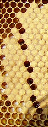
Beekeepers must be able to recognise bee diseases and parasites and to differentiate the more serious diseases from those that are less important. Because of variation in appearance it is possible to mistake chalk brood for EFB and sacbrood for AFB, or the other way round. Brood combs of healthy colonies usually exhibit a solid and compact brood pattern. Almost every cell of the comb central area will contain an egg, larva, or pupa. The cappings of sealed cells will be uniform looking and of an oatmeal colour, each capping will be convex, but not domed or bullet shaped. If you have a few cells that are vacant, but they follow the line of frame wiring, it may well be the intrusion of the wire into the base of the cell that causes the cell not to be used, but as shown in the image at right, the regularity of occurrence of such cells will be obvious to you.
In contrast brood combs of diseased colonies usually have a spotty brood pattern (pepperbox appearance that is called "shotgun brood" in USA), the cappings are often darker and concave, some may be perforated. The capped cells may be more varied in colouration as well as being generally darker. The combs may contain scales caused by the dried remains of AFB infected larvae sticking lengthwise on the lowest side of brood cells, these can be difficult to see, so face away from the light, angle the combs in order that sunlight can shine over your shoulder to illuminate the lower cell surfaces.
However... If you open a hive and you see nice even brood patterns and light oatmeal coloured cappings, do not be complacent. You still need to be vigilant and look at every cell as it is easy to be lulled into a false sense of security by the apparently good combs. I would say it is easy to miss a single diseased larva or cell under these circumstances.
Bee Inspectors will use a technique for freeing the frames of bees that I will describe, but I have a personal dislike of this method. I (D.A.C.) have seen it performed badly, with many bees damaged and indeed frames broken by heavy handed application of the process, which I found unnecessary with my own stock as they were much less excitable than the majority of bees found in UK. I (R.P.) have seen great contrasts in the handling ability of colonies of bees by Bee Inspectors, varying from being very gentle to close on as rough as I have ever seen. In the latter the Bee Inspector justified it by saying that inspecting for disease is much more invasive than a normal inspection! It seems strange that some can manage it better than others can!.
Method of examination... This text is a direct copy of instructions.
- Remove the outside comb, which is unlikely to contain brood, and lean it against a front corner of the hive. You will then have room to work.
- Take each comb in turn, and holding it by the lugs within the brood chamber, give it a sharp shake. This will deposit the bees on the bottom of the hive without harming them, the queen or brood. (In well over 50 years of beekeeping I can only remember a queen being killed or maimed on half a dozen occasions, two of which were by Bee Inspectors demonstrating shaking combs within the brood box as described above. Their argument is that bees don't fly, but if you shake the frame firmly over the brood box and the bees are good, there is no problem anyway. R.P.)
Scan the brood area of each side of each comb that contains brood in any stage, look for areas that exhibit a different colour either in cappings or in the larvae themselves. Look for evenness of capping and pay particular attention if there is a perforation in a capping. However, it should be noted that the perforations we are looking for are small and irregular, not the roughly circular, large area associated with bald brood. A further distinguishing mark for bald brood is that the edge of the hole in the capping may be raised like a very short chimney.
American Foulbrood (AFB)
American foul brood (AFB) is caused by a spore-forming bacterium called Paenibacillus larvae, subspecies larvae. A.W. Woodrow (1941) established that the larvae become infected between hatching and 24 hrs old, when they consume AFB spores in the food they are given. Larvae susceptibility decreases from that point until 2 days and 5 hours old when they become immune. The spores germinate in the gut, becoming bacteria that move into the tissues, where they multiply vigourously. The infected larvae die after their cell is sealed, and millions of infective spores are produced in the remains of their bodies. These remains dry to form scales, which stick tightly to the cell wall and often cannot be removed by the bees. Thus brood combs from infected colonies are heavily contaminated with many millions of spores.
European Foulbrood (EFB)
European foul brood (EFB) is caused by a bacterium called Melissococcus plutonius. Infected food is fed to larvae, the bacteria multiply in the mid - gut and competes with the larvae for its food. Starvation is the result and is why EFB shows in the unsealed stage, although it can also show after the cell is sealed.
Organisms associated with European foul brood
Some organisms do not cause European foul brood, but they influence the odour and consistency of the dead brood and can be helpful in diagnosis. These secondary invaders include Paenibacillus alvei (formerly Bacillus alvei).
Stonebrood
Stonebrood is a fungal disease of larva first described in 1906 by Maassen. It is said to be caused by several fungi from the genus Aspergillus, including A. flavus, A. fumigatus, A. niger. These fungi are common soil inhabitants that are also pathogenic to mammals, other insects and birds. The disease is difficult to identify in its early stages of infection. The fungus grows rapidly and forms a characteristic whitish-yellow collar-like ring near the head end of the infected larva. A wet mount prepared from the larva shows mycelia penetrating throughout the insect. After death, the infected larva becomes hardened and quite difficult to crush. Hence the name Stonebrood. Eventually, the fungus erupts from the integument of the insect and forms a false skin. At this stage, the larva may be covered with green, powdery fungal spores. The spores of A. flavus are yellow green, those of A. fumigatus are grey green, and those of A. niger, black. These spores can become so numerous that they fill the comb cells containing the affected larvae.
Stonebrood can usually be diagnosed from gross symptoms, but positive identification of the fungus requires its cultivation in the laboratory and subsequent examination of its conidial heads. Aspergillus spp. can be grown on potato dextrose or Sabouraud dextrose agars. Caution: These fungi can cause respiratory diseases in humans and other animals.
(In over 50 years of beekeeping I have never seen stonebrood, so probably not worth much attention, or certainly not in the U.K. R.P.)
Chalkbrood
Chalkbrood is a fungal disease of larva first "identified" in 1913 by Maassen. It is a common disease that does little more than hinder the growth of a colony. There are several reasons why one colony may have a higher incidence than others in the same apiary.
Protozoan Diseases
Protozoa are predominately microscopic and usually occur as single cells. No protozoa are commonly found in association with the brood of honey bees.
Nosema
There are two nosemas that affect honey bees, Nosema apis that has been with Apis mellifera probably for millions of years and Apis ceranae that has recently jumped species.
Coprological examination
By examining faecal material, Nosema can also be detected without sacrificing workers or queens. On glass plates
collect faeces of worker bees near the hive entrance, scrape off a deposit, mix it with water, and prepare
a wet mount from the resulting suspension (Wilson and Ellis 1966). Suspect queens can be confined in small petri dishes
or in glass tubes and allowed to walk freely. They usually defecate within 1 hour. Queen faeces
appear as drops of clear, colourless liquid, which can then be transferred to a microscope slide with a pipette or capillary
tube. Place a cover glass over the faeces before examination with a high, dry objective (L'Arrivee and
Hrytsak 1964). Other Protozoa Gregarines Four gregarines (protozoans of the order Gregarinida) are associated with honey bees: Monoica apis, Apigregarina
stammeri, Acuta rousseaui, Leidyana apis. Gregarines are found attached to the epithelium of the midgut of
adult honey bees. To view gregarines, gently remove the midgut from the digestive tract of a suspect bee and place it on
a microscope slide in a drop of water. The midgut can be separated from the digestive tract at the point
of attachment with the proventriculus (honey stomach) and hindgut, using fine tweezers and a scalpel. Gently break open
the midgut with fine tweezers and a probe, and place a cover glass over the resulting suspension.
Gregarines can be seen using the low-power objective of a compound microscope. Viral Diseases
Sacbrood
Sacbrood is the only common brood disease caused by a virus. Since sacbrood-diseased larvae are relatively free from bacteria, laboratory verification is usually based on gross symptoms and the absence of bacteria. Positive diagnosis requires the use of a special antiserum or molecular techniques. Affected larvae change from pearly white to grey and finally black. Death occurs when the larvae are upright, just before pupation. Consequently, affected larvae are usually found in capped cells. Head development of diseased larvae is typically retarded. The head region is usually darker than the rest of the body and may lean toward the centre of the cell. When affected larvae are carefully removed from their cells, they appear to be a sac filled with water. Typically the scales are brittle but easy to remove. Sacbrood-diseased larvae have no characteristic odour.
(When I started keeping bees in 1963 all the books mentioned sacbrood as if it was a common problem. Even when travelling to other parts I hardly saw it until about 2005, when it became quite common, to the point where I have been called out many times because beekeepers thought they had AFB, which to an inexperienced eye it can look a little bit like. For some reason it has become much more common. R.P.)
Chronic Bee Paralysis (CBPV)
Adult bees affected by chronic bee paralysis are usually found on the top bars of the combs. They appear to tremble uncontrollably and are unable to fly. In severe cases, large numbers of bees are found crawling out the hive entrance, probably ejected by healthy bees. Individual bees are frequently black, hairless, and shiny. In some cases, paralysis-like symptoms can be caused by toxic chemicals. Ideally, the diagnosis of this disease is made using serological techniques. Since this is beyond the capability of most laboratories, diagnosis is usually made by observing symptoms in individual bees and, when possible, colony behaviour.
CBPV has been much more of a problem since the introduction of varroa, probably because of it being vectored by the mite.
Filamentous Virus
Filamentous virus is also known as F-virus and bee rickettsiosis. This disease, previously thought to be of rickettsial origin, can be diagnosed by examining the hemolymph of infected adult bees using dark-field or phase-contrast microscopy. The hemolymph of infected honey bees is milky white and contains many spherical to rod-shaped viral particles of a size close to the limit of resolution for light microscopy.Acute Paralysis Bee Virus and Kashmir Bee Virus
Acute paralysis bee virus (APBV) and Kashmir bee virus (KBV) are two serologically related viruses, and the antiserum produced from one virus will cross-react with the other virus (Hung et al. 1996). These viruses commonly occur in apparently healthy adult bees. No specific gross symptoms have been attributed to either virus. Whereas APBV is a disease of adults, KBV is reported to cause mortality in brood and adult honey bees. APBV and KBV diseases can be diagnosed using immunodiffusion tests. Recently, molecular methods were developed for detecting both diseases.Parasitic Mite Syndrome (PMS)
Colonies infested with parasitic mites can display an array of symptoms referred to as the parasitic mite syndrome. The syndrome affects both adult bees and brood. It is quite likely this syndrome is similar to that reported by Ball (1988) who refers to the condition as a "secondary infection" in colonies infested with V. jacobsoni.
Acarine (Acarapis woodi). Known in the U.S. as Tracheal Mites.
Three Acarapis species are associated with adult honey bees: Acarapis woodi, A. externus, A. dorsalis. Only A. woodi is known to be harmful. All three species are difficult to detect and identify because of their small size and similarity, so they are frequently identified by location on the bee instead of by morphological characters.
Despite the fuss that is made of acarine and the probably erroneous view that it caused the Isle of Wight Disease, I have only seen problems in colonies that were headed by recently imported queens, usually Italians. In the U.S. there have been problems in the 21st century, but they have a lot of Italian bees.
Melittiphis alvearius
Melittiphis alvearius is a little-known mite that is associated with honey bees but is not considered to be a pest, not being a parasite or predator, more likely a pollen scavenger.
Disease Interactions
Paenibacillus larvae apparently produces a potent antibiotic that eliminates competition from other bacteria associated with honey bee larvae. For this reason, American foul brood (AFB) and European foul brood (EFB) are rarely found in the same colony, except in cases where AFB is just becoming established in colonies that already have EFB.
Ascosphaera apis the fungal organisem that causes Chalk Brood produces linoleic acid, which inhibits the growth of Paenibacillus larvae and Melissococcus pluton. Since the introduction of chalk brood into the United States, the incidence of EFB has fallen dramatically. However, the incidence of AFB appears to have remained constant.
It is not unusual to find chalkbrood and sacbrood on the same comb or on a comb with larvae infected with AFB, although apparently no single larva has been found to be infected with more than one disease. This is an important point to remember when selecting a sample for disease diagnosis.
Mixed infections in adult bees are more common. Adult bees could be infected with N. apis and also be infected with viruses or spiroplasma. It is also possible for adult bees to be infested with one or more species of mites.
Originally written by Dave Cushman. Edited with additions by Roger Patterson.